
Image Acquisition Methods Development
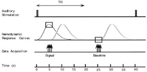
It has been the philosophy of the FNL from its inception that the examination of neuropsychiatric questions using imaging should not be limited by existing techniques. There has always therefore been an emphasis on developing new methodology, as needed, to better address specific neurobiological questions. Multiple PET and fMRI acquisition methods have been developed. Many of these techniques have been incorporated into the ongoing studies of the FNL, such as the reduction of susceptibility artifact at the base of the brain using z-shim echo planar imaging methods,silent, event-related, sparse-sampling fMRI method which avoids the confound of scanner noise (which is especially important for auditory and some psychiatric studies), and high resolution fMRI.

Processing and Analysis Methods for Functional Magnetic Resonance Imaging
In the FNL, fMRI analysis methods are continually being developed and optimized to improve detection of the small fMRI signals associated with the neuropsychological paradigms used in the MRI scanner. This work is being done with and by Dr. Hong Pan, with his trainees. Analytic techniques have been developed to improve detection power and estimation accuracy: (1) Based on variance component analysis, appropriate statistical modeling of residuals in fMRI data has been incorporated, which includes additive confounds (such as estimated head motion, physiological noise, global CBF fluctuation, scanner drift, experimental periods) and temporal correlation. (2) Based on mixed-effects statistical models, the intra- and inter-subject variabilities have been taken into account to allow results to be more generalizable to the populations studied. (3) Multi-level hierarchical mixed-effects models have been developed, with more advanced statistical modeling of the variance-mean relationship, for the multi-level nested data structures (such as trials/ runs/ sessions/ subjects/ subject groups/ study sites) and heteroscedastic variances (i.e. differing variance at different values of a random variable) common in fMRI data sets, to take advantage of and to meet the challenge presented by multi-site studies. (4) Correlation analyses of behavioral, physiological and clinical measures with fMRI data provides additional and important information about underlying pathophysiology. (5) Extraction of temporal trends in fMRI time-course provides information about changes in brain activity over the course of the experiment, which is important in understanding modulation of function. (6) Region-of-interest analyses, looking with greater specificity at the structure and function of particular brain areas (including the amygdala, hippocampus, and ventromedial prefrontal cortex), are being conducted. (7) Significant improvements have been made in existing, external software for performing voxel-based morphometry (VBM), a technique for examining differences in structure across the entire brain. (8) Multivariate statistical techniques have been employed to allow the study of interactions among brain areas, which is crucial, as we now understand that it is how brain regions function together, rather than activity in isolated areas, that is also likely to be relevant in the study of neuropsychiatric disorders. Overall, these novel approaches to characterizing and modeling many aspects of the MRI signal are resulting in improved ability to detect subtle but important changes in key brain regions. In addition to statistical and computational advances, numerous improvements in automation, visualization, and software user interfaces have been made as well.
Neuropsychological Paradigm Development, Psychophysics and Psychophysiological Measures
The FNL has significant expertise in neuropsychological paradigm development. The interpretation of functional differences detected between particular groups of subjects is largely dependent upon what neuropsychological task the subject is performing during the activation study. Task design must be targeted to probe either a brain function of interest, or particular neurocircuitry. Exquisite attention must be paid to creating appropriate control conditions to isolate the cognitive component of interest (e.g. in a subtraction paradigm), or to parametrically vary one component of the task to hone in on the corresponding brain function.
An alternative to using activation paradigms is to examine differences in resting state patterns of BOLD fMRI signal in different subject populations, which can provide complementary information. In addition, the FNL routinely uses MRI blood flow techniques to obtain resting activity measures.


Multimodal fMRI/EEG
Combining non-invasive methods with high spatial and high temporal resolution of neural activity is essential for resolving the complex neural dynamics and functional connectivity of human perceptual and cognitive networks. We use multimodal fMRI/EEG to probe neural networks with high spatiotemporal resolution. We previously reported a linear correlation in human auditory cortex between hemodynamic fMRI and mass-neural EEG measurements, supporting the idea that common neural sources contribute to these responses, as demonstrated unequivocally in invasive animal studies.
Pan H, Epstein J, Silbersweig DA, Stern E. New and emerging imaging techniques for mapping brain circuitry. Brain Res Rev. 2011 Jun 24;67(1-2):226-51. Epub 2011 Feb 24
Yang Y, Gu H, Silberweig DA, Stern E. Simultaneous perfusion and BOLD measurements using single-shot interleaved Z-shim echo-planar imaging (SSIZS-EPI). Magnetic Resonance in Medicine, 53: 1207-1211, 2005.
Pan H, Chen Q, Stern E, Silbersweig DA. Multilevel nonlinear mixed-effects models for nested factors in functional brain imaging. Proceedings of the 2003 Joint Statistical Meeting, San Francisco, CA, American Statistical Association, pp. 3162-3167.
Gu H, Feng H, Zhan W, Silbersweig DA, Stern E, Yang Y. Single-shot interleaved Z-Shim EPI with optimized compensation for signal losses due to susceptibility-induced field inhomogeneity at 3T. NeuroImage 17:1358-64, 2002.
Zhan W, Gu H, Silbersweig DA, Stern E, Yang Y. Inversion profiles of adiabatic inversion pulses for flowing spins: the effects on labeling efficiency and labeling accuracy in perfusion imaging with pulsed arterial spin-labeling. Reson. Imag, 20:487-94, 2002.
Zhan W, Gu H, Xu S, Silbersweig DA, Stern E, Yang Y. Circular spectrum mapping for intravoxel fiber structures based on on high angular resolution apparent diffusion coefficients. Reson. Med, 49: 1077-88, 2003.
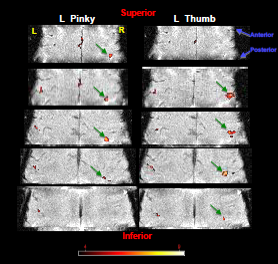
Engelien A, Yang Y, Engelien W, Zonana J, Stern E, Silbersweig DA. Physiological Mapping of Human Auditory Cortices with a Silent Event-Related fMRI Technique. Neuroimage, 16:944-953, 2002.Yang Y, Gu H, Zhan W, Xu S, Silbersweig
DA, Stern E. Simultaneous perfusion and BOLD imaging using reversed spiral scanning at 3T: characterization of functional contrasts and susceptibility artifacts. Reson. Med, 48:278-89, 2002.
Gu H, Engelien W, Feng H, Silbersweig DA, Stern E, Yang Y. Mapping transient, randomly occurring neuropsychological events using independent component analysis. Neuroimage, 14: 1432-1443, 2001
Stern E, Silbersweig DA. Symptom capture as a strategy for methodologic development and pathophysiologic investigation in functional neuropsychiatric imaging, in Strategies in psychiatric neuroimaging: clinical and research applications. Dougherty and S Rauch, eds. American Psychiatric Press, 125-142, 2001.
Yang Y, Engelien W, Pan H, Xu S, Silbersweig DA, Stern E. A CBF-based event-related brain activation paradigm: Characterization of impulse-response function and comparison to BOLD. Neuroimage, 12, 287-297, 2000.
Yang Y, Engelien W, Xu S, Silbersweig DA, Stern E. Transit time, trailing time and cerebral blood flow during brain activation: measurement using multislice, pulsed spin-labeling perfusion imaging, Magnetic Resonance in Medicine, 44:680-685, 2000.
Yang Y, Engelien A, Engelien W, Xu S, Stern E, Silbersweig DA. A Silent Event-Related Functional MRI Technique for Brain Activation Studies without Interference of Scanner Acoustic Noise. Magnetic Resonance in Medicine, 43:185-90,2000.
Jones T, Silbersweig DA, Stern E, Schnorr L, Seaward J, Clark JC, Lammertsma AA, Grootoonk S. The development of in-vivo tracer methods to obtain new information about human disease: a study of the hallucinating brain. Eur J Nucl Med , 23: 332-335, 1996.
Silbersweig D, Stern E, Frith CD, Cahill C, Schnorr L, Grootoonk S, Spinks T, Clark J, Frackowiak R, Jones T. Detection of thirty-second cognitive activations in single subjects with positron emission tomography: a new low dose H215O regional cerebral blood flow three-dimensional imaging technique. J Cereb Blood Flow Metab 13: 617-629, 1993
Silbersweig D, Stern E, Schnorr L, Frith CD, Ashburner J, Cahill C, Frackowiak R, Jones T. Imaging transient, randomly-occurring neuropsychological events in single subjects with positron emission tomography: an event-related countrate correlational analysis. J Cereb Blood Flow Metab, 14: 771-782, 1994.
Liebenthal E, Ellingson ML, Spanaki MV, Prieto TE, Ropella KM, Binder JR (2003) Simultaneous ERP and FMRI of the auditory cortex in a passive oddball paradigm. Neuroimage, 19 (4):1395-1404.
Ellingson ML, Liebenthal E, Spanaki MV, Prieto TE, Binder JR, Ropella KM (2004). Reduction of Ballistocardiogram artifact in the simultaneous acquisition of auditory event-related potentials and functional resonance images. Neuroimage, 22 (4):1534-1542.